About
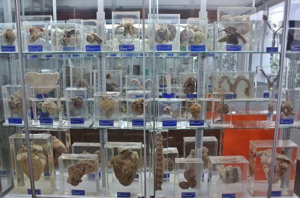
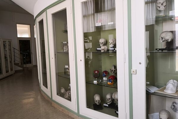
On the ground floor of the Faculty of Veterinary Medicine of the Universidad Nacional Autónoma de México (National Autonomous University of Mexico), a room with huge display cases holds hundreds of animal specimens. This is the Museo de Anatomopatología (Museum of Pathological Anatomy), an exhibit showing the macabre diseases that can affect animals.
The museum is visited by hundreds of students and professors of the biological sciences every year. Its oldest specimens date back to 1938, when the collection was started by Dr. Manuel H. Sarvide, who was then the head of the Department of Pathology. The museum moved into new facilities in 1971, and was expanded by Dr. Aline Schunemann de Aluja. In 1991, the collection was installed in its current location.
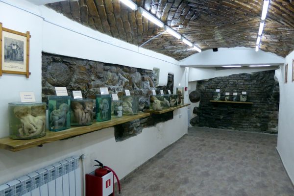
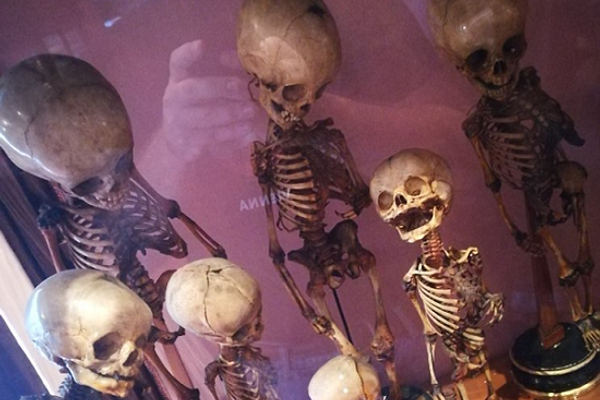
Among the collection you can find examples of liver with cirrhosis, tumors, lung infarctions, carcinoma of the eyes, and organs with cysticercosis (an infection caused by tapeworm larvae). All kinds of animals are included: Cows, bats, whales, goats, and even two marmoset fetuses. There are also a number of preserved parasites, from huge worms to tiny microorganisms.
Some of the most interesting specimens are familiar animals with deformities that they were either born with or acquired during their lives. Among these are a three-legged chicken; a cat born with one eye; a dog without a nose; and the impressive head of an Alaskan dog with a prominent tumor.
Related Tags
Know Before You Go
The entrance to the facility is located on Circuito Escolar. Once inside, there are signs indicating the location of the exhibit. The museum is open on weekdays from 10 a.m. to 4 p.m. Admission is free.
NEW - Yucatan: Astronomy, Pyramids & Mayan Legends
Mayan legends, ancient craters, lost cities, and stunning constellations.
Book NowCommunity Contributors
Added By
Published
December 20, 2019
